
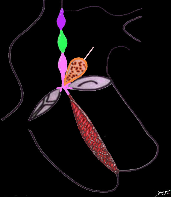
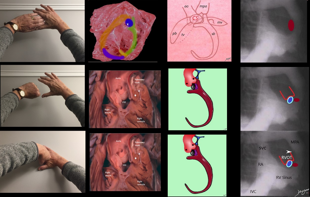
The atrial septum consists of 3 parts; The most superior is the septum derived from the sinus venosus (purple), the middle is the septum primum (green) and the most inferior is derived from the endocardial cushions (pink) The endocardial cushions also contribute to the formation of the of the membranous component of the interventricular septum. The interventricular septum consists of the membranous septum and the muscular septum (red). The great vessels are separated by a muscular conal septum (orange) and the membranous aorticopulmonary septum (light pink).
Ashley Davidoff MD TheCommonVein.net
The Conal septum had two little lambs (Ao and PA)
And everywhere the conal septum went
The sheep were sure to go
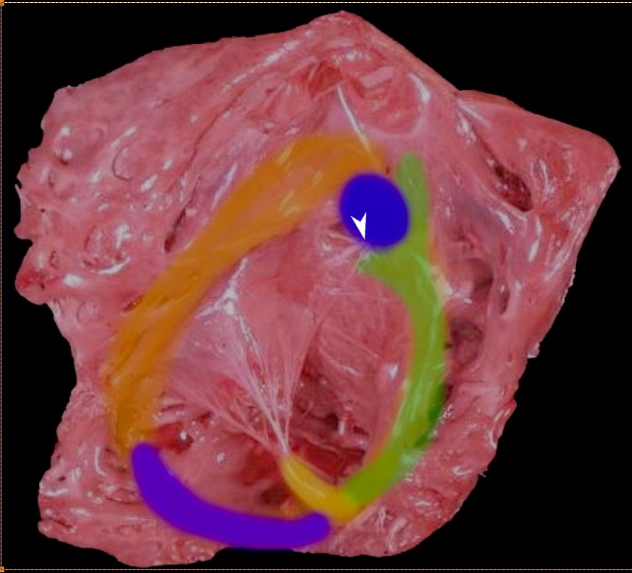
Thr superior aspect of the septal band (green) has two limbs – the “Y” of the septal band. and the conal septum (blue) embeds itself in the Y of the septal band. The (chordae) of the conal papillary muscle (aka papillary muscle of Lancisi- white arrow) also inserts in the “Y ” of the septal band. The anterolateral papillary muscle (yellow) )originates from the septal band) and subtends the anterior leaflet of the tricuspid valve . The moderator band (purple) connects the septum to the the free wall, and the parietal band (orange) completes the muscular ring that separates the inflow from the outflow tract. The pulmonary valve (above the parietal band and conal septum) defines the border between the RVOT and conus with the pulmonary artery.
Ashley Davidoff MD
06409 RV muscle bands b01.81
Ashley Davidoff MD TheCommonvein.net
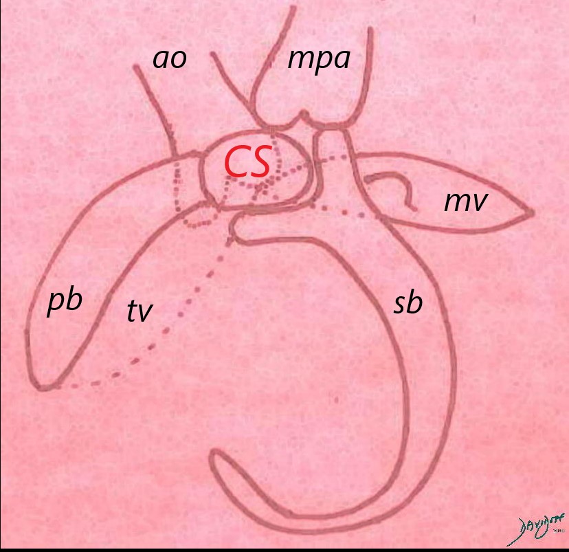
The conal septum lies in the ‘Y” of the septal band (sb) and the medial tip of the parietal band (pb). The pulmonary artery lies superior and to the left of the the conal septum and the aorta lies posterior and the conal septum. Since the conal septum lies between to the two great vessels, any change in its position affects both great vessels. If for example it moves posteriorly toward the aorta, it will cause subaortic stenosis, and if it moves leftward it will cause subpulmonary stenosis.
Ashley Davidoff MD TheCommonvein.net
normal Conal Septum in Tetralogy of Fallot (TOF) (Monology of Stensen)
In TOF the conal septum (purple) is too small resulting in a VSD, (black hole) and also misaligned toward the left and anterior causing subpulmonary infundibular stenosis and an overriding aorta. Since the aorta and PA are bound together by the conal septum – shift of the conal septum results in movement of both. The subpulmonary stenosis results in decreased forward flow and hence structures distal to the infundibulum may become hypoplastic, dysplastic or aplastic. Associated anomalies for example include absent pulmonary valve (PV) pulmonary stenosis or atresia, stenotic branch pulmonary stenosis, or absent pulmonary arteries
Ashley Davidoff MD
TheCommonVein.net
THe deficient and malaligned conal septum
Ashley Davidoff MD TheCommonVein.net
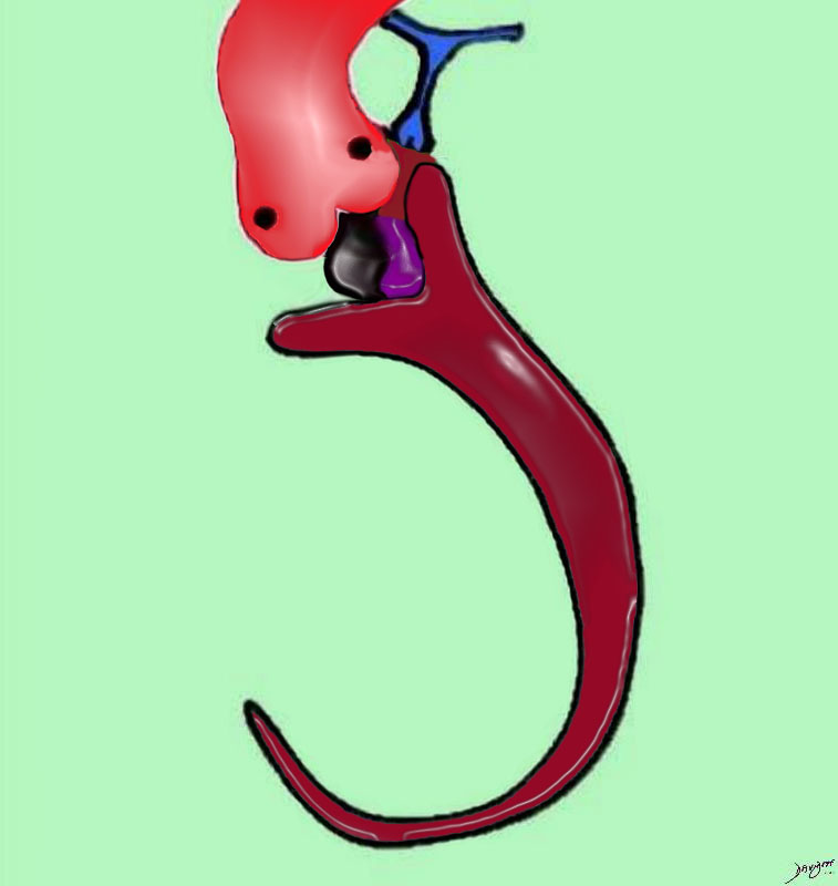
In TOF the conal septum (purple) is too small resulting in a VSD, (black hole) and also misaligned toward the left and anterior causing subpulmonary infundibular stenosis and an overriding aorta. Since the aorta and PA are bound together by the conal septum – shift of the conal septum results in movement of both. The subpulmonary stenosis results in decreased forward flow and hence structures distal to the infundibulum may become hypoplastic, dysplastic or aplastic. Associated anomalies for example include absent pulmonary valve (PV) pulmonary stenosis or atresia, stenotic branch pulmonary stenosis, or absent pulmonary arteries
Ashley Davidoff MD
TheCommonVein.net
Sotiria C. Apostolopoulou, Athanassios Manginas, Nikolaos L. Kelekis & Michel Noutsias
BMC Cardiovascular Disorders volume 19, Article number: 7 (2019)
The Common Vein.net

The RV angiogram (a, b) shows a stenotic infundibulum in the A-P projection (white arrow) and lateral projection The overriding aorta fills later through a VSD. The second series is an LV gram (c,d) with catheter entering the RA, passing through an ASD into the left atrium and then LV. The aorta is well opacified and is normal in size in contrast to the smaller main pulmonary artery, and branch pulmonary arteries
Ashley Davidoff MD TheCommonVein.net
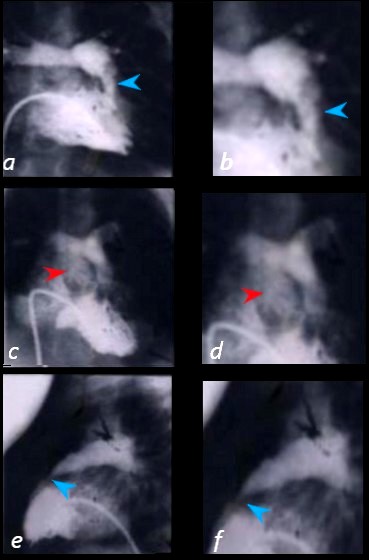
The angiogram shows a catheter entering the right atrium and RV from the IVC. Contrast injected into the RV traverses a stenotic infundibulum in the A-P projection (blue arrow) – (a -magnified in b ) and lateral projection (e and magnified in f) The overriding aorta fills later through a VSD – (red arrow – c and magnified in d)
Ashley Davidoff MD TheCommonVein.net
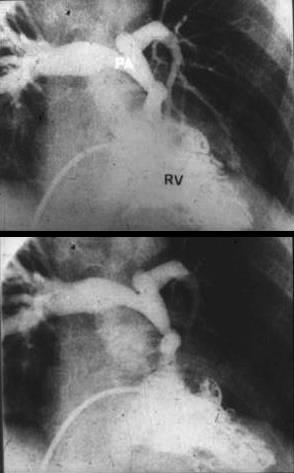
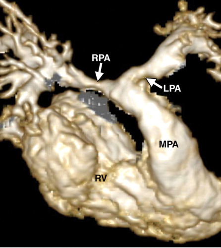
The RV angiogram in the A-P projection shows a stenotic infundibulum , dysplastic appearing pulmonary valve region, supravalvar stenosis, hypoplastic main pulmonary artery and a dilated right pulmonary artery The overriding aorta fills later through a VSD.
Ashley Davidoff MD TheCommonVein.net
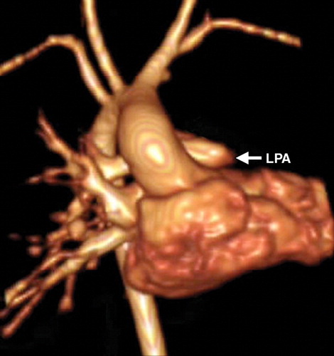
(Frank L et al Cardiovascular MR Imaging of Conotruncal Anomalies Radiographics)

(Frank L et al Cardiovascular MR Imaging of Conotruncal Anomalies Radiographics)
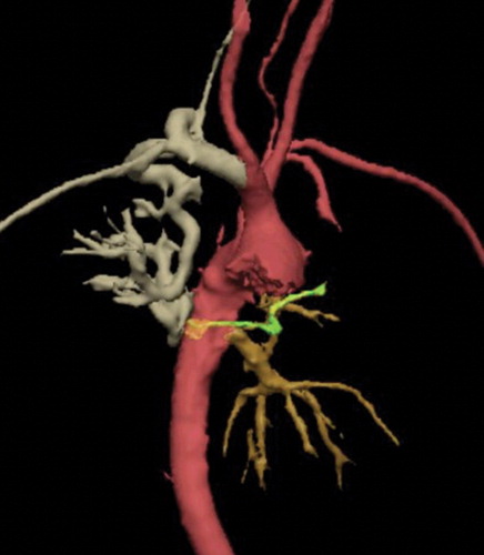
(Frank L et al Cardiovascular MR Imaging of Conotruncal Anomalies Radiographics)
(Frank L et al Cardiovascular MR Imaging of Conotruncal Anomalies Radiographics)
(Frank L et al Cardiovascular MR Imaging of Conotruncal Anomalies Radiographics)

(Frank L et al Cardiovascular MR Imaging of Conotruncal Anomalies Radiographics)

(Frank L et al Cardiovascular MR Imaging of Conotruncal Anomalies Radiographics)
(Frank L et al Cardiovascular MR Imaging of Conotruncal Anomalies Radiographics)
References
- TCV
- Conotruncal Abnormalities
- Embryology
- Anatomy of the Right Ventricle
- Double Outlet Right Ventricle
- Double Outlet Left Ventricle
- Tetralogy of Fallot
- Transposition of the Great Vessels
- Truncus Arteriosus